Water‑clear cell parathyroid adenoma: A case report and mini‑review of the literature
- Authors:
- Published online on: September 17, 2025 https://doi.org/10.3892/mi.2025.269
- Article Number: 70
-
Copyright : © Abdullah et al. This is an open access article distributed under the terms of Creative Commons Attribution License [CC BY 4.0].
Metrics:
Total
Views: 0 (Spandidos Publications: | PMC Statistics:
)
Total PDF Downloads: 0 (Spandidos Publications: | PMC Statistics:
)
Abstract
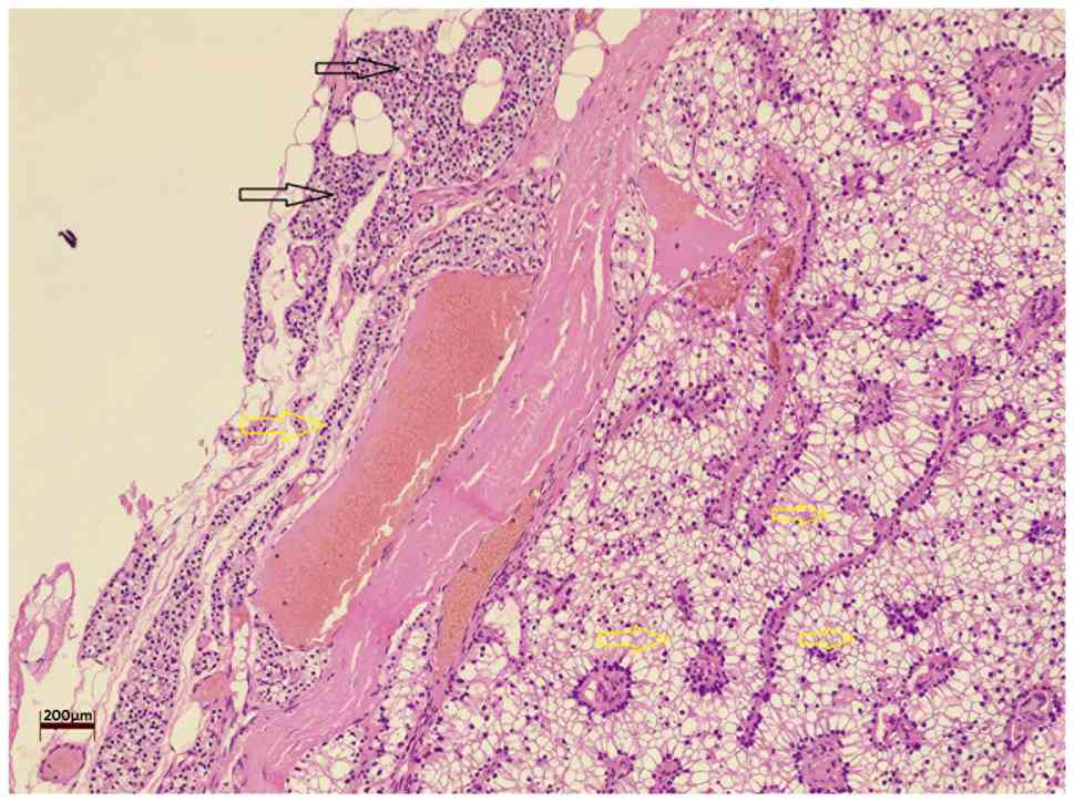
Water‑clear cell parathyroid adenoma (WCCPA) is a rare variant of parathyroid adenoma, characterized by large polygonal cells with optically clear cytoplasm. To date, only a limited number of cases have been reported in the English literature. The present study describes the case of a patient with WCCPA. An 80‑year‑old female presented with muscle pain and fatigue lasting for 1 week. A clinical examination revealed a normal thyroid gland with no palpable masses. Laboratory tests revealed elevated levels of serum calcium (12.73 mg/dl) and parathyroid hormone (PTH; 303 pg/ml). A neck ultrasonography revealed a 28x16x11 mm hypoechoic, mildly vascular lesion, suspicious of a parathyroid nodule, located posterior to the left thyroid lobe. A left lower parathyroidectomy was performed, and a histopathological examination confirmed the diagnosis of WCCPA. Post‑operatively, the serum calcium and PTH levels of the patient normalized, and her recovery was uneventful. In addition, a review of 4 recent cases of WCCPA found in the literature demonstrated a common clinical pattern of elevated calcium levels and increased PTH levels. Imaging techniques, such as ultrasonography and sestamibi scans revealed localized parathyroid lesions, which were subsequently confirmed as WCCPA through histopathological evaluation. Surgical excision was performed in all cases, resulting in postoperative normalization of calcium levels in the majority of patients. WCCPA is a rare, yet significant cause of primary hyperparathyroidism. An accurate diagnosis relies on a histopathological examination, as clinical and imaging findings may be non‑specific. Surgery can yield favorable outcomes.